We are accepting new patients. Book an Appointment NOW!

A clinical physical therapy expert with over 30+ years of experience in rehabilitation and patient care. A graduate of Utkal University, he specializes in orthopedic, neurological, and vestibular therapy. Passionate about compassionate, evidence-based treatment, he is dedicated to helping patients restore mobility, reduce pain, and regain independence.
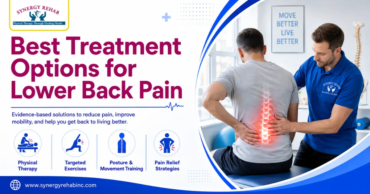
Lower back pain treatment should start with the reason pain began, not with a random exercise or painkiller. A recent strain, a stiff
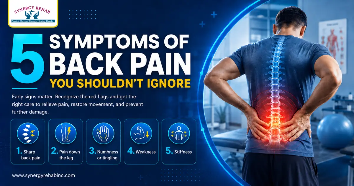
Back pain is common, but some pain patterns should not be treated like a normal muscle strain. The most important symptoms of back
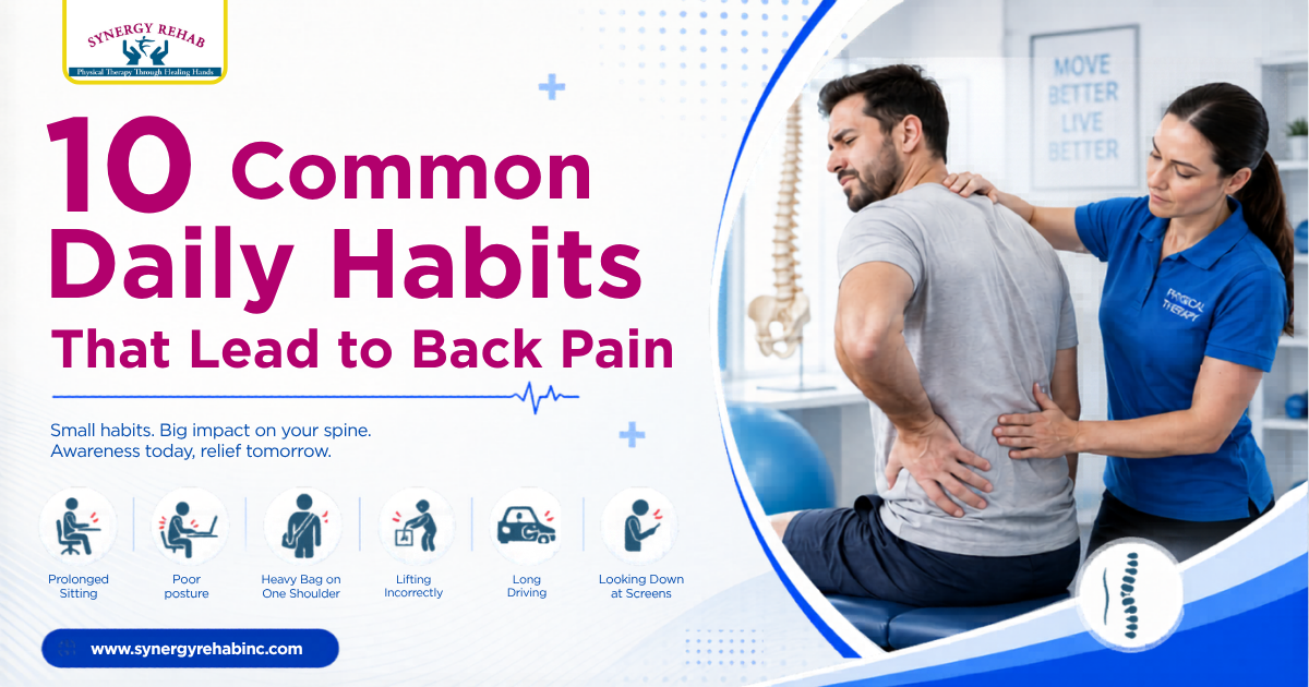
Back pain can build slowly from repeated daily stress, not only from one sudden injury. Many people notice stiffness after long sitting, poor
Upper back pain can make sitting, reaching, driving, sleeping, or deep breathing uncomfortable. Pain may appear between the shoulder blades, near the neck,

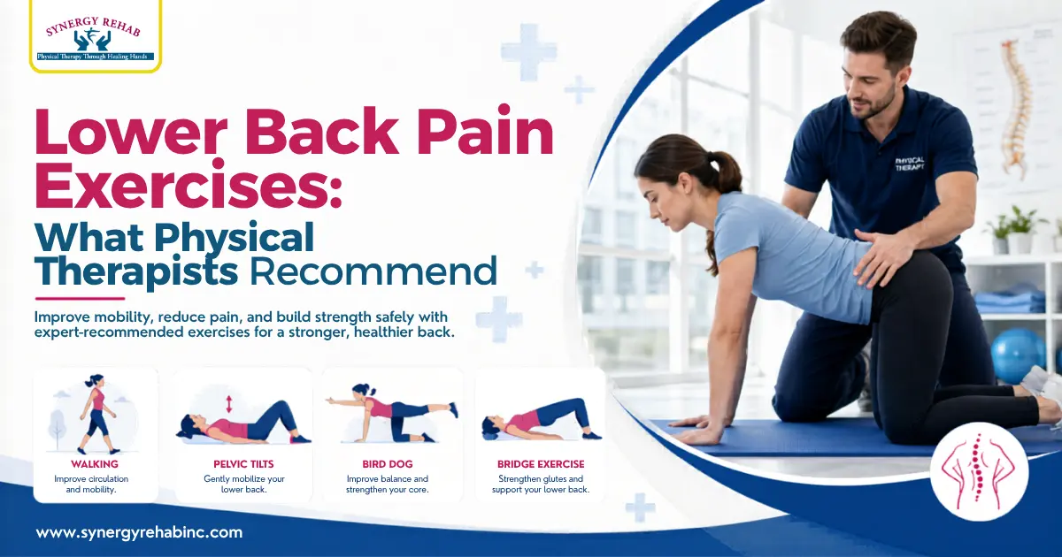
Lower back pain exercises can help improve mobility, reduce stiffness, and rebuild strength, but the safest plan depends on what is causing your
Understanding the cost of physical therapy for back pain helps patients plan care before starting treatment. The price may vary based on symptoms,

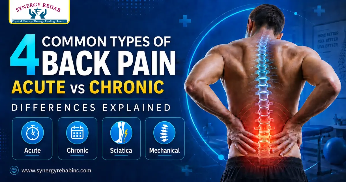
Back pain is not one single problem. The main types of back pain can be grouped by how long pain lasts, where it
Back Pain Treatment should start by finding what is stressing your spine, muscles, joints, or nerves. For many people in Southfield, pain may

Back pain is not just a symptom. It often reflects how your body moves, rests, and responds to daily stress. In clinical practice,
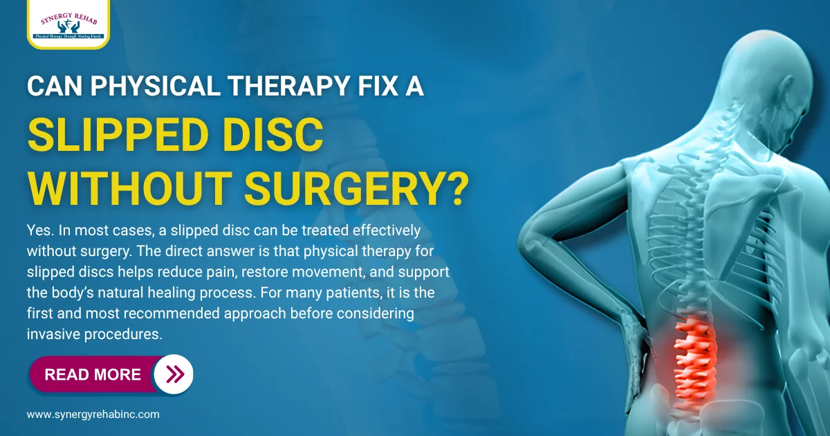
Yes. In most cases, a slipped disc can be treated effectively without surgery. The direct answer is that physical therapy for slipped discs